


The New Lens of Medicine: How AI is Transforming Medical Imaging
Written by Leonardo Viola
Medical image analysis is currently undergoing one of its most transformative eras, where artificial intelligence is no longer a future ambition, but a real tool to increase diagnostic accuracy and optimize the work of specialists.
Those who follow medical research have hardly escaped the wave of studies claiming that artificial intelligence can match, or even surpass, radiologists in interpreting medical images. And the numbers are truly impressive. Vision Transformers, being one of the most established and best-performing architectures, achieve over 99% accuracy in retinal exams and in detecting COVID-19. Ensemble models, which consist of combining various AI models, score above 90% in key diagnostic benchmarks. AI can detect osteoarthritis in X-rays up to three years before the patient experiences any symptoms.
These data show that this is not just about marginal improvements, but a real shift in what automated analysis is capable of. The progress spans various modalities, from chest X-rays and CT scans to MRIs, histopathology slides, and ultrasounds, and is achieved through a growing variety of architectures: large-scale foundational models, models capable of generating synthetic medical images for training, and even experimental architectures inspired by quantum computing.
QuantumSpace developments will transform image analysis in the biomedical sector
How it works
To better understand the impact and scope of these tools in the clinical environment, it is important to first briefly analyze how these models process information. Deep learning systems applied to diagnostic imaging are usually structured in a three-phase process. Initially, the medical image is introduced into the neural network, which is responsible for extracting its fundamental visual characteristics. Next, the network identifies the regions of interest and predicts the respective clinical labels, ideally accompanied by bounding boxes and confidence scores. Finally, post-processing algorithms overlay these detections onto the original image to generate the final diagnosis and the respective confidence index.
The central objective of these tools is to act as a decision support system for radiologists, and never as a substitute for human judgment. For this support to be truly effective in practice, the technical excellence of the model must translate into robust clinical results, something that is clearly reflected in the accuracy metrics that these algorithms have been achieving.
Beyond pure diagnosis, algorithms are also beginning to take on the cognitive load that slows down clinical workflows. Automated reporting tools can translate imaging findings into structured radiology reports, associating observations with specific image regions. Generative models create synthetic training images that reduce radiation exposure and bypass patient privacy restrictions. Federated learning frameworks allow dozens of hospitals to collaboratively train a model without ever sharing a single patient file, ensuring anonymity and confidentiality. We are not talking about futuristic concepts, but about recent published and peer-reviewed literature, where several are already being evaluated in a clinical context.
Our technologies will support clinicians and medical teams in making informed decisions based on structured data
What are the steps back?
However, the same literature that celebrates these results is surprisingly frank when it comes to their limitations. The central problem is not accuracy, which is already guaranteed to some extent, but generalization. Almost all high-performing models were trained and tested with data from large academic hospitals in the West. When these models encounter patients from different demographics, geographies, and/or imaging equipment, performance degrades significantly. One study found that the United States alone contributes more publicly available imaging datasets than all of Africa, Latin America, South Asia, and Southeast Asia combined. A model with 90% overall accuracy may only perform at 70% in elderly or minority patients.
Data is the common thread running through almost all the limitations encountered in the field. Data sets are very small, very homogeneous, and difficult to share for legal reasons.
Another frequently mentioned difficulty is the high computational complexity of some models. In the context of medical diagnosis, these systems require not only significant processing power but also access to a significantly larger volume of clinical data and medical images to ensure the robustness and reliability of the results. However, this technological demand clashes with the reality of the sector, since not all institutions have the financial capacity or the necessary infrastructure to acquire and maintain computers with this processing capacity.
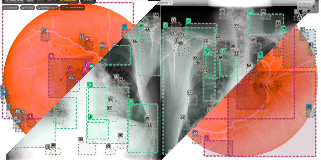
Another extremely relevant issue is the interpretability, which proves insufficient to generate clinical confidence. This is the main reason why an entire subfield of Explainable AI, called XAI, has emerged. It uses heat maps and attention mechanisms to show clinicians which regions of the image influenced the presented prediction.
Current regulatory frameworks have not kept pace with this rate of technological innovation. Laws such as HIPAA and GDPR were created before the emergence of concepts like synthetic data or federated learning, which have generated a legal vacuum that technology alone cannot fill. Even the most promising approaches, such as quantum machine learning, are still in the pre-clinical phase. Currently, their benefits are minimal and limited to laboratory simulations or studies with limited data in the field of oncology.
What emerges from the literature, considered as a whole, is an area that is technically ahead of its institutional infrastructure. The algorithms are real, the accuracy gains are real, and the potential to reduce the workload of overburdened radiologists and clinicians is real. But the path between benchmark performance and routine clinical use involves interpretability, equitable construction of datasets, regulatory modernization, and rigorous real-world validation, none of which can be solved simply with better architecture.
The technology developed at QuantumSpace will fill the gap between algorithmic potential and practical reality
QuantumSpace and Lens
It is precisely to fill this gap between algorithmic potential and practical reality that QuantumSpace emerges. As a visual intelligence platform that combines Artificial Intelligence, Computer Vision, and Data Science, QuantumSpace focuses on transforming unstructured images into measurable and comparable knowledge, aligning technological innovation with the most rigorous best practices in data governance.
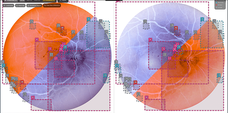
QuantumSpace’s direct response to this regulatory and clinical challenge is embodied in Lens: the explainable comparator for medical images, designed to highlight significant differences and support traceable clinical review.
Specifically designed to highlight significant visual differences and support a fully traceable clinical review, Lens is designed to be anything but a “black box.” By focusing on automatic standardization, visual detection, clear explainability, and a secure and transparent history, the software translates the potential of AI into a safe, real-world tool, ensuring that the final decision always remains where it should be: in the physician’s clinical judgment.
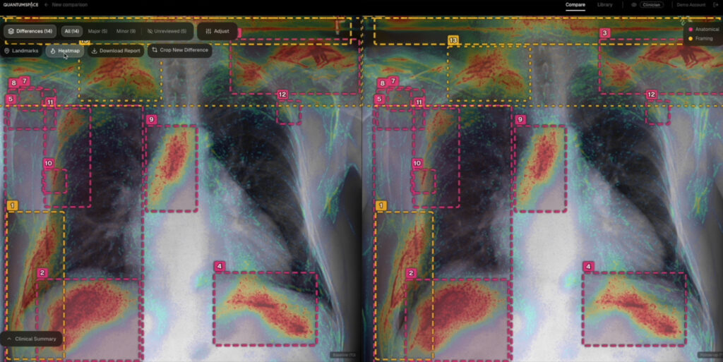
The Lens system optimizes the analysis of imaging exams over time through a structured three-phase workflow. Initially, the software performs technical normalization of the images, automatically correcting variations in format, scale, resolution, and orientation to ensure that comparisons reflect only real anatomical changes. Then, through dynamic and explainable analysis, the platform uses medically tailored visual models to segment areas of interest, calculate evolution metrics, and generate clear textual explanations associated with each visual alert.
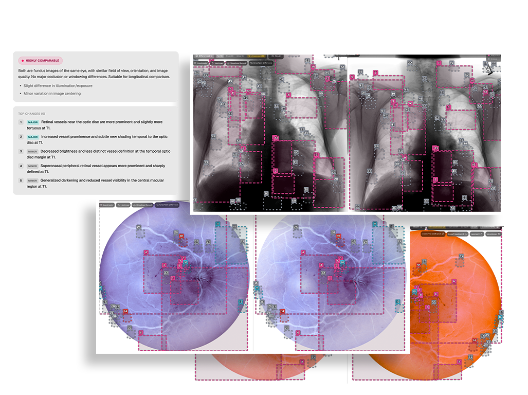
Finally, to ensure traceability and process security, the system rejects the opacity of the “black box” by recording all notes and decisions in an inviolable history, an essential step to facilitate reporting, audits, and secure collaboration between medical institutions. Lens optimizes physicians’ workflow in seconds, transforming complex visual data into a clear, transparent, and auditable chronological evolution.
Our projects paves the way for a path toward a more intelligent and connected future for biomedical innovation
A clear path
The analysis of biomedical images through artificial intelligence is at an unprecedented point of technical maturity: models exist, they work, and in controlled contexts, they often exceed expectations. But the real challenge has never been strictly technological. It is institutional, ethical, and human: building datasets that represent the full diversity of patients, developing models that clinicians can understand and trust, and creating regulatory frameworks capable of keeping pace with innovation.
The transition from the laboratory to the clinic requires not only better algorithms, but also deeper collaboration between engineers, physicians, regulators, and patients. When this convergence happens, AI will not replace clinical judgment; it will amplify it, making healthcare faster, more equitable, and more accurate for everyone.
References
Aburass, S., Dorgham, O., Al Shaqsi, J., Abu Rumman, M., & Al-Kadi, O. (2025). Vision Transformers in Medical Imaging: a Comprehensive Review of Advancements and Applications Across Multiple Diseases. In Journal of Imaging Informatics in Medicine (Vol. 38, Number 6, pp. 3928–3971). Springer Nature. https://doi.org/10.1007/s10278-025-01481-y
Afnouch, M., Bougourzi, F., Gaddour, O., Dornaika, F., & Ahmed, A. T. (2025). Artificial intelligence in bone metastasis analysis: Current advancements, opportunities and challenges. In Computers in Biology and Medicine (Vol. 194). Elsevier Ltd. https://doi.org/10.1016/j.compbiomed.2025.110372
Ahmed, F., Naz, N. S., Khan, S., Rehman, A. U., Ismael, W. M., & Khan, M. A. (2026). Explainable artificial intelligence (XAI) in medical imaging: a systematic review of techniques, applications, and challenges. BMC Medical Imaging, 26(1). https://doi.org/10.1186/s12880-025-02118-w
Alshanbari, A. H., & Alzahrani, S. M. (2025). Generative AI for Diagnostic Medical Imaging: A Review. Current Medical Imaging Formerly Current Medical Imaging Reviews, 21. https://doi.org/10.2174/0115734056369157250212095252
Assaf, R., Rammal, A., Goupil, A., Kacim, M., & Vrabie, V. (2025). Topological data analysis and machine learning for COVID-19 detection in CT scan lung images. BMC Biomedical Engineering, 7(1). https://doi.org/10.1186/s42490-025-00089-1
Fang, M., Wang, Z., Pan, S., Feng, X., Zhao, Y., Hou, D., Wu, L., Xie, X., Zhang, X. Y., Tian, J., & Dong, D. (2025). Large models in medical imaging: Advances and prospects. In Chinese Medical Journal (Vol. 138, Number 14, pp. 1647–1664). Lippincott Williams and Wilkins. https://doi.org/10.1097/CM9.0000000000003699
Filano, R., Gde, I., Dirgayussa, E., Alfarabi, A., Lailatul Akbar, R., & Zakiah, H. (2025). Advancements and Challenges of Deep Learning in Diagnostic Radiology: A Systematic Literature Review Article Info. Jurnal Fisika, 15(2), 38–50. https://journal.unnes.ac.id/nju/index.php/jf/index
Ghosh, D., Mehjabin, M., Rayed, M. E., Mridha, M. F., & Kabir, M. M. (2026). Advancements and challenges of federated learning in medical imaging: a systematic literature review. Artificial Intelligence Review, 59(2). https://doi.org/10.1007/s10462-025-11489-z
Koetzier, L. R., Wu, J., Mastrodicasa, D., Lutz, A., Chung, M., Koszek, W. A., Pratap, J., Chaudhari, A. S., Rajpurkar, P., Lungren, M. P., & Willemink, M. J. (2024). Generating Synthetic Data for Medical Imaging. In Radiology (Vol. 312, Number 3). Radiological Society of North America Inc. https://doi.org/10.1148/radiol.232471
Liu, J., Cen, X., Yi, C., Wang, F. A., Ding, J., Cheng, J., Wu, Q., Gai, B., Zhou, Y., He, R., Gao, F., & Li, Y. (2025). Challenges in AI-driven Biomedical Multimodal Data Fusion and Analysis. In Genomics, Proteomics and Bioinformatics (Vol. 23, Number 1). Beijing Genomics Institute. https://doi.org/10.1093/gpbjnl/qzaf011
Musa, A., Prasad, R., Onwualu, P., & Hernandez, M. (2026). A Systematic Review of Cross-Population Shifts in Medical Imaging Analysis with Deep Learning. Big Data and Cognitive Computing, 10(3), 76. https://doi.org/10.3390/bdcc10030076
Oettl, F. C., Zsidai, B., Oeding, J. F., Hirschmann, M. T., Feldt, R., Fendrich, D., Kraeutler, M. J., Winkler, P. W., Szaro, P., & Samuelsson, K. (2025). Artificial intelligence-assisted analysis of musculoskeletal imaging—A narrative review of the current state of machine learning models. Knee Surgery, Sports Traumatology, Arthroscopy, 33(8), 3032–3038. https://doi.org/10.1002/ksa.12702
Supriyadi, M. R., Samah, A. B. A., Muliadi, J., Awang, R. A. R., Ismail, N. H., Majid, H. A., Othman, M. S. Bin, & Hashim, S. Z. B. M. (2025). A systematic literature review: exploring the challenges of ensemble model for medical imaging. In BMC Medical Imaging (Vol. 25, Number 1). BioMed Central Ltd. https://doi.org/10.1186/s12880-025-01667-4
Related Posts:

QuantumSpace x Sykes Tech: the next layer of Biomedical Intelligence
QuantumSpace's new strategic partnership with Sykes Tech represents a concrete step toward building an applicable, scalable, and relevant technology in the biomedical sector. Explore with us the true potential of this joint operation.
QuantumSpace at AI Week Milan 2026: Expanding Data Intelligence
The report of QuantumSpace's participation at AI Week 2026, the most prominent AI event in Europe. Confronting with the general public, our CEO and IT Lead Developer showcase the potential of our patented technologies.
What Is Visual Intelligence? Building the Infrastructure of the Visual Economy
This article will introduce you to QuantumSpace's Vision as a growing Company in a challenging market. Discover how we face the challenges in multiple areas of research, spearheading developments in the fields of Quantum and AI.